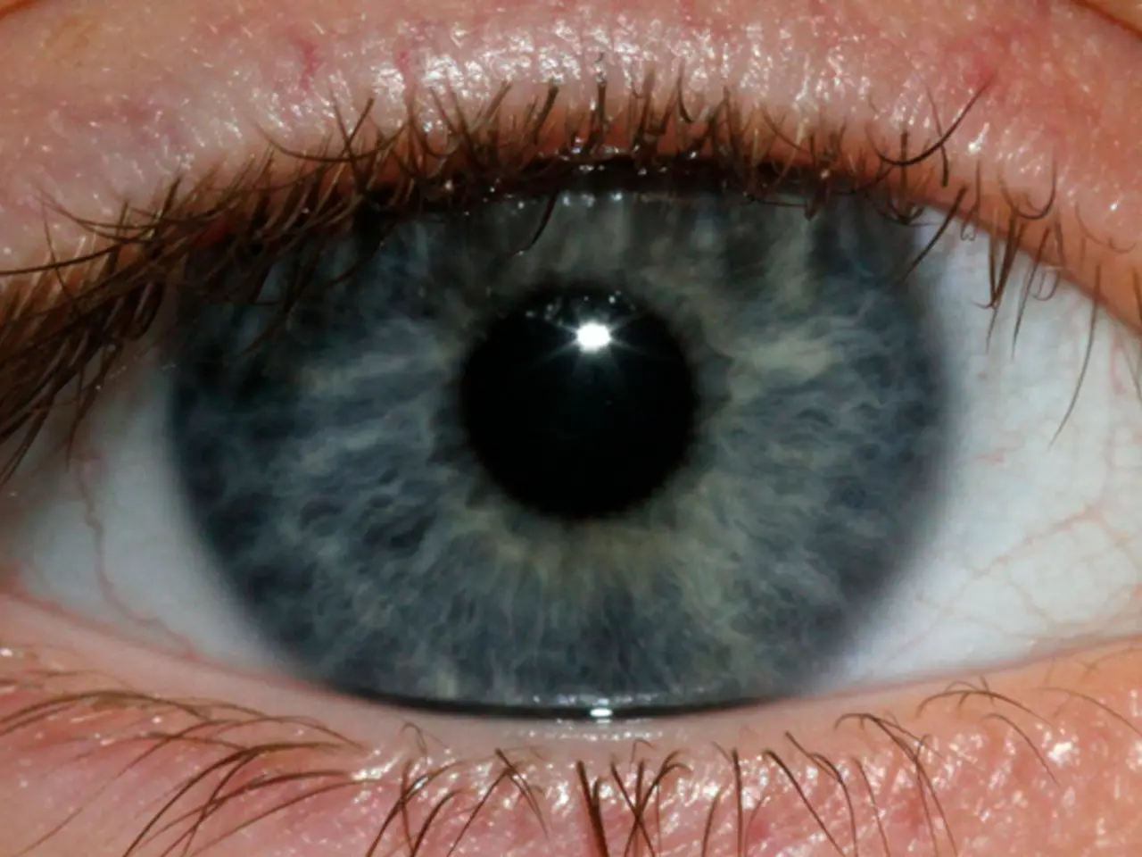
Anatomical exploration of the eyeball
In an engaging science activity designed for students, they are given the opportunity to delve into the intricacies of the eye through an eye dissection. This activity, using a preserved cow or sheep eye specimen, allows students to examine the parts of an eyeball up close.
Materials Required
- Preserved cow or sheep eye (cow eyes are often used due to their large size, making anatomy easier to observe)
- Dissection kit including scissors, scalpel, forceps, and dissecting tray
- Gloves and personal protective equipment (PPE)
- Student guide with labeled images of eye anatomy
- Teacher's manual with instructions and additional resources
- Waste disposal materials (for cleaning up after dissection)
Steps for the Dissection
- Preparation: Put on PPE and arrange your dissection tools and specimen on the dissection tray.
- External examination: Identify external features such as the cornea, sclera, and optic nerve.
- Cutting the eye: Hold the eye firmly and use scissors or a scalpel to cut the eye in half, separating the front and back halves.
- Internal observation: Remove the vitreous humor (gel-like substance inside the eye) and extract the lens, noting its hardness due to preservation.
- Dissect the cornea: Cut around the cornea's edge to expose the iris and pupil.
- Remove the iris: Use forceps to carefully detach the iris from the sclera to study its structure.
- Examine retina and optic nerve: Observe the retina attached to the back and its connection to the optic nerve, noting the blind spot.
- Inspect tapetum lucidum: If present (in cow eyes), look for this reflective layer beneath the retina.
- Cleanup: Dispose of specimen parts properly and clean all tools and surfaces.
The dissection guide usually includes identification and function of about 13 major parts, such as the cornea, iris, pupil, ciliary muscle, sclera, vitreous humor, retina, blind spot, choroid, optic nerve, tapetum lucidum, lens, and aqueous humor.
This activity can be completed in 1–2 class periods and aids students in learning about mammalian eye anatomy and function through hands-on observation. Some commercial kits come with all supplies and instructions needed except PPE.
Additional digital resources and 3D models may be available to support teaching and understanding of the eye's complex structure and physiology. The Labelling the eye activity, published by Referencing Hub media, is another related activity that allows students to name key components of an eye and learn about and identify parts of the human eye.
Dissections are a powerful method to engage students in learning about life processes, and this eye dissection activity does not require any prior demonstration, video, or expert visit. By the end of the activity, students should be able to link the parts of an eyeball to their functions, enhancing their understanding of sight, depth perception, and the complexities of our vision.
For those interested in further exploring human vision, resources such as A related activity to further explore the human eye or Ophthalmology research can provide additional information. A Word file for the eye dissection activity can be downloaded, but the link was not provided in the given information.
- The exploration of medical-conditions specific to eye-health, such as understanding the complexities of vision, can be expanded with activities like the Labelling the eye activity, which requires students to name key components of both the cow and human eye.
- This dissection activity, along with additional resources like A related activity to further explore the human eye, can help students gain a deeper understanding of different health-and-wellness aspects related to the eye, even leading them to pursue ophthalmology research.

